Bacterium excluded from the Eukaryote Club
It’s something you learn in high school – there are two basic approaches to cellular life – prokaryotes (bacteria and archaea) and eukaryotes (the rest of us – aardvarks, amoebae, apricots, etc.). Prokaryotes have an open-plan office, with all biological functions carried out in the one cellular space. Eukaryotes, however, have dedicated compartments for the chief executive (nucleus), finance (mitochondria), sales and marketing (golgi, endoplasmic reticulum), and so on.
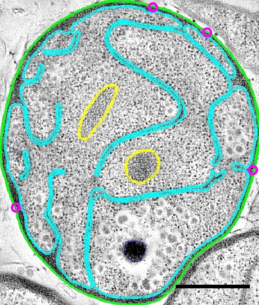
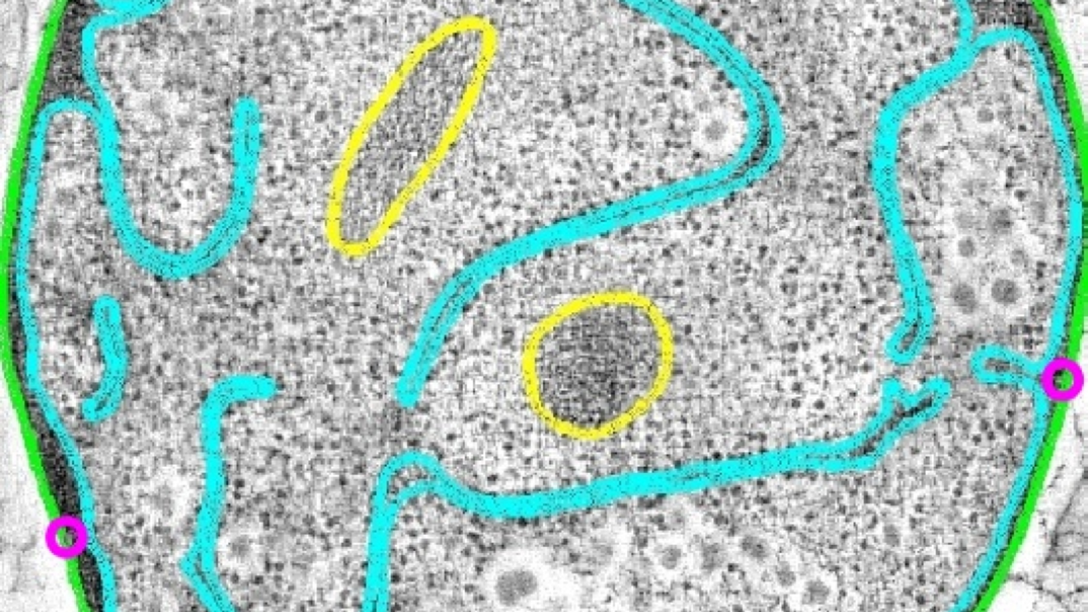
But every now and then you get an upstart prokaryote that seems to have ideas above its station. One such is Gemmata obscuriglobus, an exemplar of a bunch of unusual bugs known as the PVC superphylum (for Planctomycetes, Verrucomicrobiae, Chlamydiae). Gemmata has a complex membrane structure, and previous studies of its 3D configuration have suggested that this bacterium has a compartmentalised cell, eukaryote-style, its genetic material encapsulated in a nucleus-like body.
With a nice combination of technical wizardry and sheer hard work, Rachel Santarella-Mellwig, Damien Devos and colleagues, authors of a paper just published in PLOS Biology, have managed to reconstruct the structure of the internal membranes of a typical Gemmata cell in spectacular detail. They embedded ten bugs in plastic, chopped them each into ten or so slices, and then took electron microscope snapshots of the Gemmata salami. In the absence of software that could do the job, they then manually tracked and assigned the membranes in each slice, building up a detailed 3D model of the membranes and other features.
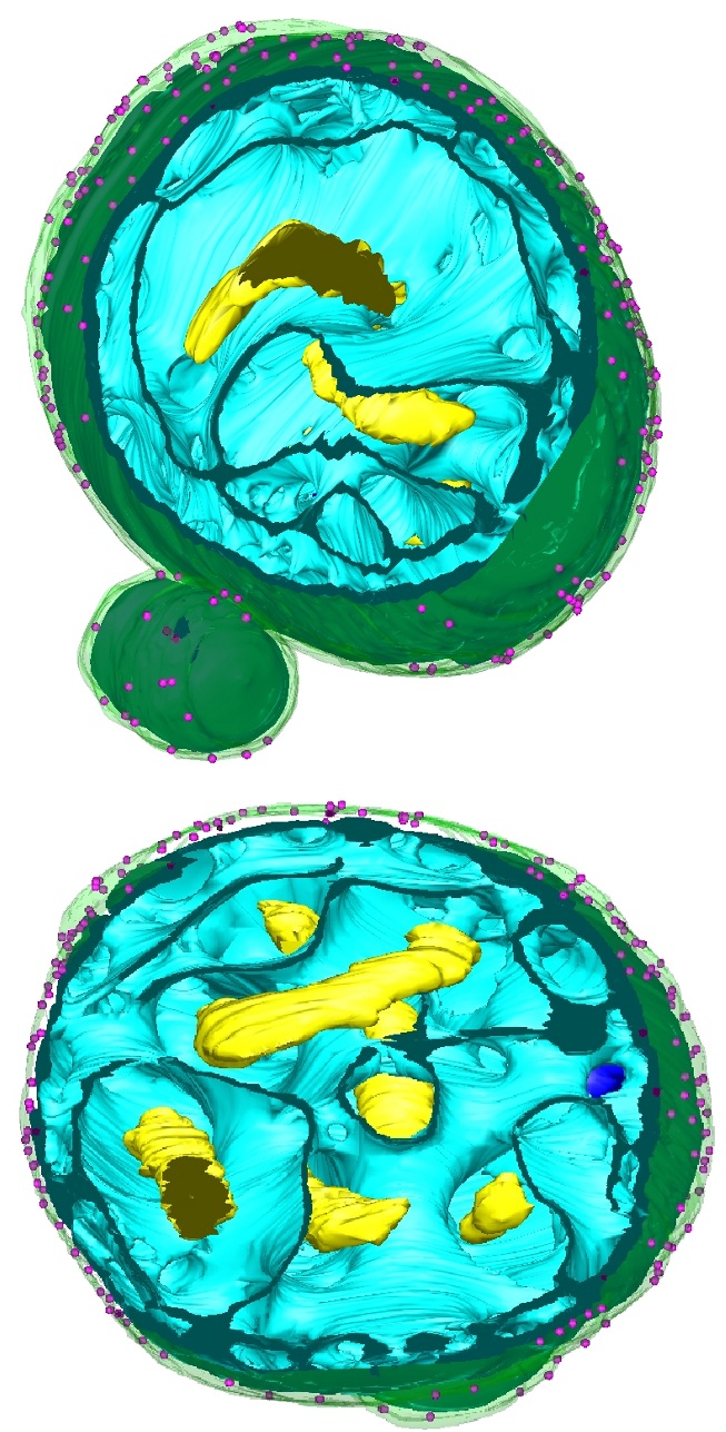
The answer, perhaps sadly, is that Gemmata is actually rather a traditional bug, topologically speaking. The beautiful pictures and movies produced by the authors reveal a membrane system that, while extremely convoluted, doesn’t enclose any separate compartments. Gemmata is as prokaryotic as they come.
The authors propose that a common ancestor of the PVC superphylum evolved a way to fold its membrane up. But what’s the point of this? They draw the comparison with lab workhorse and tabloid horror story, Escherichia coli – the archetypal bug body-plan. Both Gemmata and E. coli are what’s called “gram-negative” bugs, in that they have two membranes – one outside the cell wall, and one inside it – separated by the periplasmic space. As an aside, it had been thought that Gemmata might lack an outer membrane – this study clearly shows that it does have one after all, and that the cytoplasm and the periplasm are the only two topologically closed compartments.
Gemmata is a 2-micron sphere. E. coli is a rod-shaped thing about 1.5 microns long. In each case the periplasmic space – the gap between the membranes – accounts for about a third of the cell’s total volume. However, the difference really shows up when you look at membrane surface area – in the classic bug E. coli the inner and outer membranes have almost identical surface areas, but the convolutions of the Gemmata inner membrane give it three times the area of the outer membrane.
The purpose of this extra membrane surface area is obscure, but the authors draw parallels with the eukaryotic endoplasmic reticulum and speculate that Gemmata and other PVC members might use it for the synthesis of sterols. Sterols are complex chemicals used by eukaryotes for signalling purposes and to modify the physical properties of membranes. However, Gemmata and its relatives are thought to be among the few bacteria capable of making sterols, and the authors imply that the association with a fancy membrane structure may not be a coincidence.
Despite the disappointing lack of full-blown compartmentalisation, there are striking similarities between the fancy internal membrane of the PVC bacteria and the endoplasmic reticulum of eukaryotes. Are the PVCs an evolutionary link between classic gram negative bugs and us, or have these common membrane features (high surface area, sterol synthesis, decoration with ribosomes) arisen twice in evolution?

Santarella-Mellwig, R., Pruggnaller, S., Roos, N., Mattaj, I., & Devos, D. (2013). Three-Dimensional Reconstruction of Bacteria with a Complex Endomembrane System PLoS Biology, 11 (5) DOI: 10.1371/journal.pbio.1001565